FLUG Webinar Series January 2025

FLUG Webinar Series January 2025

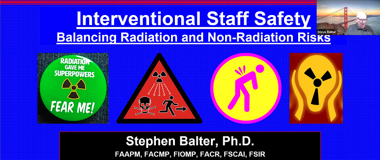
Weekly throughout January 2025 FLUG will be running a webinar series covering a range of topical issues related to the use of fluoroscopy in medicine. Attendance is free, though there will be a small charge for a certificate of attendance if required. Thursday 9th January – 14:00 UTC+0 Staff Radiation Safety: Balancing radiation & non-radiation risksProfessor Steve Balter[Professor of Clinical Radiology, Columbia University Medical School, NewYork City] Register now Thursday 16th January – 14:00 UTC+0 Results from the BSIR survey of UK Interventional Radiology radiationprotection practiceDr. Bella Hausen[Consultant Interventional Radiologist, Lancashire Teaching Hospitals] Register now POSTPONED Thursday 23th January – 14:00 UTC+0 This webinar has been postponed. Further details to follow. 1.Radiographer advanced practice in Uroradiology interventionsObinna Nwankwo[Advanced Uroradiology Practitioner, Interventional Radiology Theatres,Nottingham University Hospitals]2.Nottingham’s vascular access service: an evolutionKimberley Croome[Advanced Radiology Vascular Practitioner, Interventional RadiologyTheatres, Nottingham University Hospitals] ...
Read More
FLUG 2024 – Single room CT Hepatic Angiography for microwave liver ablation imaging – Start of a new  service; Ed McDonagh

FLUG 2024 – Single room CT Hepatic Angiography for microwave liver ablation imaging – Start of a new service; Ed McDonagh

1500-McDonaghFLUG2024CTHA-1Download Ed McDonagh, Louise Giansante, Jodie Basso, Shaira Farooq, Nicos Fotiadis, EW Johnston Purpose: Contrast-enhanced CT scans are routinely acquired before and after microwave liver ablation for planning and assessing the relationship between tumours and ablation zones, i.e. treatment efficacy. However, delivering contrast through peripherally inserted intravenous cannulas limits the number of acquisitions to a maximum of three scans, due to iodinated contrast dose. CT hepatic angiography involves positioning an arterial catheter, usually in the common or proper hepatic artery for selective contrast administration. This both reduces contrast dose and improves image quality for tumour and ablation zone depiction prior to, during and after ablation. Multiple contrast enhanced scans may be performed for improved ablation zone assessment and for on-table ablation zone extension if necessary in the same anaesthetic session. Placement of the catheter requires ultrasound and fluoroscopy guidance. The X-ray angiography suite and the interventional CT scanner are not co-located at our institution, and therefore placing the catheter in the Angiography...
Read More
FLUG 2024 – Occupational dose reduction: Using a floor mounted mobile lead radiation protection shield; Hannah Scott-Rees

FLUG 2024 – Occupational dose reduction: Using a floor mounted mobile lead radiation protection shield; Hannah Scott-Rees

With the increasing number and complexity of interventional cardiology procedures there is the potential of higher occupational radiation doses to interventionists. Significant development has been achieved with mobile lead equivalent radiation protection devices, providing enhanced radiation protection without the requirement of being directly worn by staff. The RAMPART M1128 radiation protection shield is one of these devices. The dose reduction provided to staff within a Cardiac Catheterisation Laboratory was assessed via use of Electronic Personal Dosimeters (EPD) with the Philips live dosimetry system DoseAware (Philips DoseAware). A 60% dose reduction to the primary operator can be achieved with the Rampart device. With further dose reductions possible for other individuals in the range of 65 to 84%. Additionally, dose rate measurements were taken in a simulated clinical set up using a phantom, which showed that the device provided a 65% dose reduction at eye level and 90% dose reduction at chest level for the primary operator position. This shows a potential...
Read More
FLUG 2024 – Trend analysis of radiation incidents from fluoroscopic or interventional examinations by healthcare providers in the British Isles; Timothy Lloyd

FLUG 2024 – Trend analysis of radiation incidents from fluoroscopic or interventional examinations by healthcare providers in the British Isles; Timothy Lloyd

The England health authority publish annual IRMER reports (CQC, 2022) using only incidents of significance (SAUE). This study considers radiation incidents by healthcare providers in several UK nations and a crown-dependency. As most radiation incidents are non-reportable to the appropriate authority, analysis is limited to SAUE.  This study considers all radiation incidents from reports made to an appointed MPE, including incidents non-reportable to authorities and analysis has been undertaken of those incidents relating to fluoroscopic and interventional examinations. A database was created from 2883 reports of radiation incidents, provided by over 100 healthcare providers to their appointed MPE, during years 2020 – 2024 with further analysis to be undertaken on 2024 data. Each radiation incident was coded using a UK coding taxonomy (RCR, 2019), and the data for each code field was analysed quantitatively. There were 52 radiation incidents relating to fluoroscopy and relating to interventional procedures. Analysis of radiation incidents has been completed detailing: reportable incidents, examination details, duty holders and...
Read More
FLUG 2024 – Reviewing the current alert level for high skin dose procedures in interventional radiology using Dosewatch; Roshni Senthilkumar

FLUG 2024 – Reviewing the current alert level for high skin dose procedures in interventional radiology using Dosewatch; Roshni Senthilkumar

Purpose: According to the Ionizing Radiation (Medical Exposure) Regulations 2017 Reg 12, the radiation dose during all procedures should be kept as low as reasonably practicable [1], however, high dose procedures are unavoidable and can lead to deterministic risk if occurred. According to the NCRP168 [2] and ICRP 2000 [3], deterministic effects can occur at a Peak Skin dose (PSD) of 2Gy or greater and medical follow-up is required. The dose area product (DAP) is the alert indicator used to estimate (PSD). The aim was to review the current DAP alert level utilized in the most common interventional radiology (IR) examinations at UHCW, with particular interest in exams for which the alert level revision was requested. A few exams had frequently been triggering the alert level even though the skin dose remained below the threshold for deterministic effects and this is identified in this report. Methods: PSD was collected using DoseWatch which is installed in all GE interventional radiology suites. DoseWatch...
Read More
FLUG 2024 – Image quality to dose optimisation in fluoroscopy using light output method – a feasibility study; Roshan S Livingstone

FLUG 2024 – Image quality to dose optimisation in fluoroscopy using light output method – a feasibility study; Roshan S Livingstone

Background: The purpose of this study was to verify the feasibility of using the light output from the display monitor to monitor contrast to background ratio (CBR) and optimise image quality in a fluoroscopy system Methods: A standard grey scale pattern (GS2 Leeds test tool) was used to assess image quality for 2 mobile Siemens Cios C-arm units (one with image intensifier and the other with a Flat panel detector (FPD – Cios Flow)). This test tool was placed into a stack of polymethyl methacrylate (PMMA) phantoms simulating a patient equivalent thickness of 10 and 20cm. Fluoroscopic images for the extremity protocol were acquired using low, medium and high dose settings for 7.5pps and continuous modes. The display monitors were checked for quality using the SMPTE pattern before assessing for luminescence (cd/m2) using an X2 Unfors light meter. The incident air kerma rate (Ka,r) at interventional reference point was recorded from the displayed values from each c-arm unit. The luminescence values...
Read More
FLUG 2024 – Developing objective measures for assessing clinical image quality in interventional radiology; Poppy Turner

FLUG 2024 – Developing objective measures for assessing clinical image quality in interventional radiology; Poppy Turner

Diagnosis and interventional procedures in healthcare rely heavily on x-ray imaging.However, there are known patient risks associated with the use of x-rays. Therefore,minimising radiation dose to the patient is essential, whilst also retaining adequate clinicalimage quality. Image quality is currently measured using test objects, which cannot bedirectly related to clinical image quality. These measures are also subjective, timeconsuming, and cannot easily be applied to large datasets. The majority of previous researchinto clinical image quality has incorporated quantitative measures of computed tomographyimage quality, with additional work aiming to automate these measures. Preliminaryresearch using quantitative measures of clinical image quality has also been carried out ininterventional radiology and digital mammography. The aim of this study was to develop objective image quality measures for interventionalradiology procedures, to be applied to large numbers of clinical images for use alongsidepatient dose data during system optimisation. This involved a retrospective audit of clinicalimages from Royal Free Hospital, and a phantom study to validate the results. The developedmetrics...
Read More